
Fujifilm: Simplifying ESD. Advancing Endoscopy.
First conceptually described almost 30 years ago, ESD is now firmly established as a feasible and effective procedure for treatment of early gastrointestinal tract (GIT) neoplasia, transforming the disease management paradigm for multiple premalignant conditions and several types of early esophageal, gastric, and colon cancers.
Despite its many advantages over other resection techniques, ESD has been slow to enter clinical practice in the West due to its technical complexity, limited existing infrastructure, and a lack of training opportunities. The development of new endoscopic technology is an ever-evolving landscape, and the transition from concept to clinical use can take years to complete. Through a process of mutual support, clinical providers and industry innovators continue to identify unmet needs in clinical practice and deliver technological enhancements aimed at improving patient care. Industry plays a key role in the development of tools that can accelerate ESD implementation and adoption, providing the necessary financial, design, and regulatory guidance.
With a longstanding commitment to innovation, Fujifilm has been a key contributor to advancing ESD technology — from its inception to more recent technological innovations — delivering more than a dozen state-of-the-art solutions in a variety of advanced endoscopic and emerging techniques.
About ESD
Endoscopic submucosal dissection (ESD) provides minimally invasive curative treatment for early-stage cancerous lesions within the gastrointestinal tract.
ESD evolved from conventional endoscopic mucosal resection (EMR) to enable en bloc resection of large, challenging lesions. Although EMR can achieve en bloc specimens for relatively small (<20 mm) lesions, most lesions with advanced pathology tend to be larger, requiring a piecemeal approach with demonstrated high rates of incomplete resection and recurrence. ESD accomplishes a slightly deeper division in the submucosa, achieving high en bloc resection rates regardless of lesion size, and facilitating precise and definitive histological assessment and staging for proper oncology follow-up. If curative, it can obviate surgery that would otherwise be needed for resection.
Video Segment:
Hiroyuki Aihara, MD, PhD
Source:
ESD has become the standard of care for various GIT precancers and early cancers in Japan and throughout East Asia, where gastric cancer prevalence is 8–10-fold higher than that of Western countries, and excellent long-term patient outcomes from high-volume centers have been reported. Mounting evidence supporting the efficacy and safety of ESD in esophageal and, more recently, colorectal applications, offers the promise of increased adoption in Europe and the United States, where colonic lesions are the most commonly encountered.
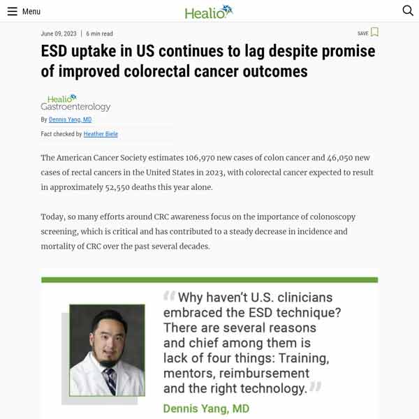
With ~ 110,000 new cases of colorectal cancer (CRC) estimated per year and > 50,000 deaths expected to result from CRC in 2023 in the US alone, arming clinicians with tools for earlier detection and more effective, efficient, and safe intervention is critical. The role of ESD in Western endoscopic practice is therefore likely to expand, calling for strategic efforts to increase awareness, training, funding, and setting or updating guidelines.
Video Segment:
Dennis Yang, MD
Peter Draganov, MD
Source:
Japanese and European guidelines endorse ESD in cases of high suspicion for submucosal invasion, especially for lesions > 20 mm, residual/recurrent carcinoma after endoscopic resection, or for lesions that cannot be optimally and radically removed by EMR because of submucosal fibrosis or chronic inflammation. In the United States, the American Society for Gastrointestinal Endoscopy (ASGE) recently published guidelines suggesting ESD over EMR for patients with early-stage, well-differentiated, nonulcerated cancer >15 mm or >20 mm for early esophageal squamous cell carcinoma (ESCC) and esophageal adenocarcinoma (EAC) management, respectively. For gastric adenocarcinoma (GAC), ASGE guidelines recommend ESD for early-stage, well or moderately differentiated, nonulcerated intestinal type cancer measuring 20–30 mm.
Major European and American societies have also released recommendations focusing on ESD application in Western populations. The European Society of Gastrointestinal Endoscopy (ESGE) recommendations advise ESD for colorectal lesions with depressed morphology and irregular or nongranular surface pattern, particularly if the lesions are larger than 20 mm. The American Gastroenterology Association (AGA) clinical practice update released in 2019 recommends ESD for colonic lesions with Kudo V-type pit pattern, depressed component (Paris 0–IIc), complex morphology (0–Is or 0–IIa+Is), rectosigmoid location, nongranular LST (adenomas) 20 mm in size, granular LST (adenomas) > or =30 mm in size, and residual or recurrent colorectal adenomas.
Video Segment:
Dennis Yang, MD
Peter Draganov, MD
Source:
What’s Advancing ESD?
Contributing factors to slower adoption of ESD procedures in the West include the lack of proper tools to facilitate visualization, retraction, and tissue manipulation — aspects that could eliminate procedure variables and simplify the overall process.
Although current endoscopic imaging allows for high-resolution images of incredible magnification, clarity, and detail, problems still arise in difficult anatomy where tight corners or folds may obscure some targets. New more-flexible scope designs, wider-angle lenses, and multiple-camera arrangements address some of these limitations. The use of hood/caps and reduplicative submucosal injection have also led to advancements in visualization. However, the supporting capacity of the transparent cap is limited, making it difficult to obtain sufficient tension in the dissection plane, while repeated injections can prolong procedure time, increasing the risk of adverse events.
Inspired by the surgical pull and push techniques, traction techniques have emerged as a functional “second-hand” while performing ESD. Traction provides adequate tissue tension and stabilization, enhancing visualization of the submucosal tissue and vasculature, and resulting in improved efficiency, safety, and outcomes.


Fig. 1, 2: The relative difficulty of ESD varies by location, with access to colon flexures, ileocecal valve/appendiceal areas, and small intestine representing the most challenging and reserved for true experts. SB, small bowel; EGJ, esophagogastric junction. (Adapted from Friedel et al. 2018, World J Gastrointest Endosc).
Video Segment:
Vivek Kumbhari, MD, PhD
Source:
Video Segment:
Dennis Yang, MD
Source:
Video Segment:
Hiroyuki Aihara, MD, PhD
Source:
Video Segment:
Hiroyuki Aihara, MD, PhD
Source:
TRACMOTION: Single operator, 360° rotatable retraction revolutionizes ESD procedures and improves patient outcomes
Although many traction methods have been developed, traction direction is limited in most, and movement of the scope can be restricted by the traction system itself. Fujifilm’s through-the-scope, single operator TRACMOTION device is designed for greater control and maneuverability, facilitating enhanced visualization and simplifying the lesion excision process:
- 360° rotatable jaws enable easy grasping and re-grasping of large lesions.
- Simple, intuitive, single-hand movements control the distal end independent of the endoscope’s movement and without the need for locking mechanisms.
- Retroflexion capabilities enhance visualization in challenging anatomy.
- Compatibility with the Fujifilm EI-740D/S Dual Channel Endoscope, as well as other dual channel endoscopes that have one 3.7mm (or larger) instrument channel and a working length of 1030mm, reduces the amount of lifting agent required, the number of device exchanges, and procedure times.

Video:
Meir Mizrahi MD, FASGE
Vivek Kumbhari, MD, PhD
Video:
Gregory Haber, MD
Video Segment:
Hiroyuki Aihara, MD, PhD
Source:
Clinical Benefits, Challenges, and Solutions
Traction techniques reduce procedure times and risk of serious adverse events
The advent of traction methods has significantly impacted ESD practice and improved patient outcomes. Traction-assisted ESD allows for unparalleled endoscopic view and tissue stability, reducing the incidence of intraprocedural bleeding or perforation, two of the major complications of ESD.
For patients, this translates to reduced table and recovery time, improved chances of curative resection, and better outcomes. In addition, the decreased technical difficulty afforded by traction techniques allows clinicians to perform higher volume of procedures with less fatigue and more confidence.
Video Segment:
Vivek Kumbhari, MD, PhD
Source:
Video:
Alexander Schlachterman, MD
Flattening the learning curve
Given these clinical benefits, what is blocking ESD from entering center stage clinical practice in the West? One of the main hurdles to overcome is related to the procedural degree of technical difficulty: ESD is more complex, technically challenging, and time- and resource-intensive, requiring extensive training and expertise. For trainees, performing ESD entails a steeper learning curve, particularly in the Western world.
In Japan and other Eastern countries, ESD training follows a traditional apprenticeship model. Formalized education is followed by observation, training in animal models, and subsequent participation during ESD cases. Training then progresses to performing cases under proctorship, beginning with smaller lesions in the distal stomach before progressing to more challenging cases in other organs.
In the West, the lower incidence of gastric cancer relative to colon and esophageal pathology is a particular challenge, limiting opportunities for hands-on training in easier to remove lesions on which beginners may develop their skills. The relative paucity in gastric cases, coupled with lack of infrastructure, dedicated devices, or structured payer reimbursement, also translate to limited numbers of endoscopists with ESD proficiency that can serve as mentors, further hindering training.
As ESD continues to gain traction in Western endoscopic practice, alternative educational models have begun to emerge. National GI societies and specialized tertiary centers offer dedicated workshops for introductory exposure to beginners, as well as more in-depth courses for those that have already started ESD in their own practice. The device and endoscope industry has also played a key role in training endoscopists, providing support and equipment for courses held by GI societies, academic institutions, and other endoscopy groups. Increased demand has led to the introduction of structured training in advanced endoscopy fellowships for select programs throughout the country, with the expectation that they may serve as a blueprint for additional programs in the future.

TRACMOTION: closing the learning gap
As an intuitive retraction tool, TRACMOTION may enable endoscopists to reduce their time in both learning and performing ESD procedures.
Video Segment:
Dennis Yang, MD
Source:
Video Segment:
Abhishek Agnihotri, MD
Alexander Schlachterman, MD
Neil Sharma, MD, FACG, FASGE, AGAF
Source:
For more information on Fujifilm’s ESD solutions, follow this link.
References
Rashid MU, Alomari M, Afraz S, Erim T. 2022. EMR and ESD: Indications, techniques and results. Surg Oncol 43:101742, PMID: 35370049, https://doi.org/10.1016/j.suronc.2022.101742.
Nishizawa T, Yahagi N. 2017. Endoscopic mucosal resection and endoscopic submucosal dissection: technique and new directions. Curr Opin Gastroenterol 33(5):315-319, PMID: 28704212, https://doi.org/10.1097/MOG.0000000000000388.
Thiruvengadam SS, Fung BM, Barakat MT, Tabibian JH. 2022. Endoscopic Mucosal Resection: Best Practices for Gastrointestinal Endoscopists. Gastroenterol Hepatol 18(3):133-144, PMID: 35506001.
Lim XC, Nistala KRY, Ng CH, Lin SY, Tan DJH, et al. 2021. Endoscopic submucosal dissection vs endoscopic mucosal resection for colorectal polyps: A meta-analysis and meta-regression with single arm analysis. World J Gastroenterol 27(25):3925-39, PMID: 34321855, https://doi.org/10.3748/wjg.v27.i25.3925.
Rotermund C, Djinbachian R, Taghiakbari M, Enderle MD, Eickhoff A, von Renteln D. 2022. Recurrence rates after endoscopic resection of large colorectal polyps: A systematic review and meta-analysis. World J Gastroenterol 28(29):4007-4018, PMID: 36157546, https://doi.org/10.3748/wjg.v28.i29.4007.
Back MK, Moon HS, Kwon IS, Park JH, Kim JS, et al. 2020. Analysis of factors associated with local recurrence after endoscopic resection of gastric epithelial dysplasia: a retrospective study. BMC Gastroenterol 20(1):148, PMID: 32397967, https://doi.org/10.1186/s12876-020-01293-0.
Bray F, Ferlay J, Soerjomataram I, Siegel RL, Torre LA, Jemal A. 2018. Global cancer statistics 2018: GLOBOCAN estimates of incidence and mortality worldwide for 36 cancers in 185 countries. CA Cancer J Clin 68(6):394-424, PMID: 30207593, https://doi.org/10.3322/caac.21492. Erratum in: CA Cancer J Clin 70(4):313.
GBD 2019 Colorectal Cancer Collaborators. 2022. Global, regional, and national burden of colorectal cancer and its risk factors, 1990-2019: a systematic analysis for the Global Burden of Disease Study 2019. Lancet Gastroenterol Hepatol 7(7):627-647, PMID: 35397795, https://doi.org/10.1016/S2468-1253(22)00044-9. Erratum in: Lancet Gastroenterol Hepatol 7(8):704.
Keihanian T, Othman MO. 2021. Colorectal Endoscopic Submucosal Dissection: An update on best practice. Clin Exp Gastroenterol 14:317-330, PMID: 34377006, https://doi.org/10.2147/CEG.S249869.
Friedel D, Stavropoulos SN. 2018. Introduction of endoscopic submucosal dissection in the West. World J Gastrointest Endosc 10(10):225-238, PMID: 30364783, https://doi.org/10.4253/wjge.v10.i10.225.
Khan S, Ali FS, Ullah S, Huang X, Li H. 2022. Advancing endoscopic traction techniques in endoscopic submucosal dissection. Front Oncol 12:1059636, PMID: 36387236, https://doi.org/10.3389/fonc.2022.1059636.
Nagata M. 2022. Advances in traction methods for endoscopic submucosal dissection: What is the best traction method and traction direction? World J Gastroenterol 28(1):1-22, PMID: 35125817, https://doi.org/10.3748/wjg.v28.i1.1.
Imaeda H, Iwao Y, Ogata H, Ichikawa H, Mori M, Hosoe N, Masaoka T, Nakashita M, Suzuki H, Inoue N, Aiura K, Nagata H, Kumai K, Hibi T. A new technique for endoscopic submucosal dissection for early gastric cancer using an external grasping forceps. Endoscopy. 2006 Oct;38(10):1007-10. doi: 10.1055/s-2006-925264. Epub 2006 Apr 27. PMID: 16673308.
Mönkemüller K, Vargo JJ. 2022. Are we ready to embrace endoscopic submucosal dissection as the organ-sparing, minimally invasive endoscopic surgical procedure of choice for large colorectal adenomas and early cancers? Gastroenterology 163(5):1168-1170, PMID: 36067819, https://doi.org/10.1053/j.gastro.2022.08.046.
Schlachterman A, Yang D, Goddard A, Gotoda T, Draganov PV. 2018. Perspectives on endoscopic submucosal dissection training in the United States: a survey analysis. Endosc Int Open 6(4):E399-E409, PMID: 29607391, https://doi.org/10.1055/s-0044-101452.
Martin JA, Gostout CJ. 2022. Evolving role of GI societies and industry in training endoscopists to perform new techniques. In: Successful Training in Gastrointestinal Endoscopy, Cohen J (Ed). Available at: https://doi.org/10.1002/9781119529675.ch38.